Focusing on professional tools and solutions
for traumatic wound repair
Integrated R&D & Precision Manufacturing for Advanced Wound Care —
exclusively serving global B2B distributors and healthcare partners.
Chongqing Qingshi Biotechnology Co., Ltd. is a China-based high-tech medical device manufacturer exclusively focused on wound care. We provide a complete product portfolio from negative-pressure drainage (VSD) to wound repair dressings and post-operative eye protection — forming a fully integrated wound care solution matrix.
Unlike simple OEM/contract manufacturers, Qingshi Bio is a technology-driven R&D enterprise. Located in Hechuan District, Chongqing, we have established long-term technical partnerships with leading universities and renowned Class-A tertiary hospitals both domestically and internationally. This collaboration has enabled us to build a closed-loop platform spanning R&D, production, and commercialization — with proprietary technology at its core.
All our core products hold NMPA Class II medical device registration certificates (Yu Xie Zhu Zhum), ensuring full compliance with regulatory standards for safety, efficacy, and traceability. Our quality management system is advancing toward ISO13485 certification.
Our entire product line revolves around a single core mission: wound care. From drainage treatment → wound repair dressings → post-operative care, we deliver end-to-end solutions across the complete wound management continuum.
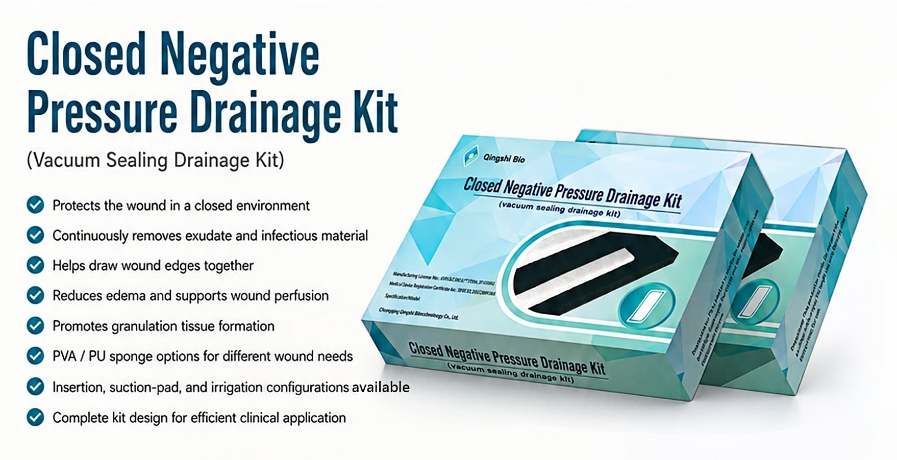
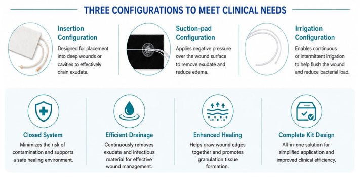
Vacuum Sealing Drainage (VSD) applies controlled negative pressure to create a sealed wound environment — efficiently draining exudate, controlling infection, accelerating granulation tissue growth, and significantly shortening healing time. Qingshi Bio's VSD platform delivers a complete, sterile, clinically-ready medical device solution.
Design Principles
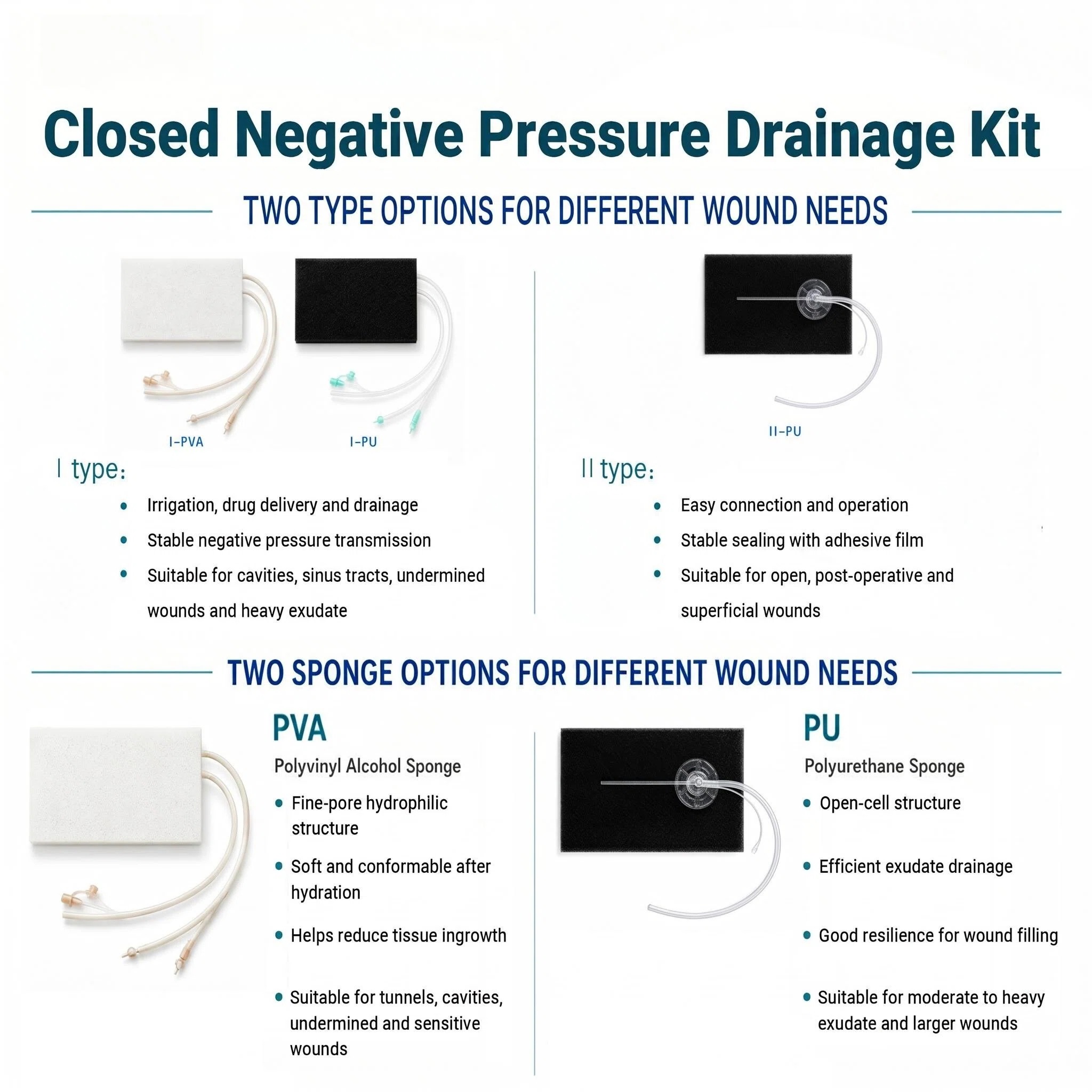
PVA Foam — Hydrophilic Material
PU Foam — Hydrophobic Material
Flow Rate ≥8 L/min — Efficient exudate removal from high-output wound beds
⚠ Contraindications
Active bleeding · Coagulopathy · Untreated osteomyelitis · Malignant tumor in wound bed
Clinical Notes
Anti-infective combo · Monitor protein/electrolytes · −125 mmHg continuous · Replace 48–72h
Clinical Indications — 10 Major Applications
1. Diabetic Foot Ulcers
VSD negative pressure promotes granulation tissue growth, effectively managing exudate and infection in diabetic foot ulcers — a critical solution for wounds that fail to heal under conventional therapy.
2. Pressure Injury (Bedsore)
Continuous negative pressure draws wound edges together, controls bioburden, and creates a moist healing environment — significantly accelerating recovery in deep pressure injuries across all stages.
3. Venous Leg Ulcers
VSD effectively manages chronic venous insufficiency-related ulcers by reducing edema, promoting circulation, and accelerating granulation tissue formation in long-standing wounds.
4. Acute Traumatic Wounds
For acute traumatic wounds requiring fast closure, VSD provides immediate negative pressure stabilization, reduces edema rapidly, and promotes quick tissue approximation — ideal for post-traumatic wound management.
5. Chronic Traumatic Wounds
For chronic traumatic wounds with prolonged healing cycles, VSD reduces edema, promotes perfusion, and stimulates proliferation — cutting healing time by up to 50% versus conventional dressing methods.
6. Burn Wounds
VSD provides effective exudate management for burn wounds, reduces infection risk, and promotes rapid granulation tissue formation — essential for second-degree and deep burn wound care.
7. Post-operative Wounds
VSD prevents surgical site infections (SSI), controls post-surgical fluid accumulation, and reduces adhesion formation risk in abdominal and thoracic procedures — accelerating post-operative recovery.
8. Surgical Site Infections
VSD effectively manages infected surgical wounds by continuous drainage of infected exudate, controlling bioburden, and promoting healthy granulation tissue formation for delayed primary closure.
9. Plastic Surgery & Dermatology
VSD is ideal for skin graft donor sites, flap procedures, and cosmetic surgery wound management — ensuring optimal aesthetic outcomes with minimal scarring and rapid healing.
10. Military & Emergency Trauma
VSD provides rapid wound stabilization for combat injuries, emergency trauma, and mass casualty scenarios — enabling field-deployable negative pressure therapy for critical wound management.
Key Warnings: Maintain continuous negative pressure for effective drainage — interruptions reduce therapeutic efficacy · Combine with anti-infective therapy (including anaerobic coverage) · Monitor protein/electrolyte loss in high-output wounds · Do not use on actively bleeding wounds or patients with severe coagulation disorders · Replace dressing when foam becomes hard, blocked, or saturated — do not exceed 72 hours
01Step 1 — Wound Preparation
Assess — Evaluate wound location, size, depth, and condition. Identify exposed bone, tendons, or vital structures.
Debride — Remove all necrotic tissue (black/brown), devitalized slough, fibrin deposits, and pus using surgical forceps, scissors, or a curette.
Irrigate — Flush the entire wound cavity with 500–1000 mL of normal saline using a syringe until effluent runs clear.
Haemostasis — Control active bleeding with pressure, electrocautery, or haemostatic agents as required.
Measure — Record wound length, width, and depth to select the correct VSD foam size.
02Step 2 — Foam Preparation & Fitting
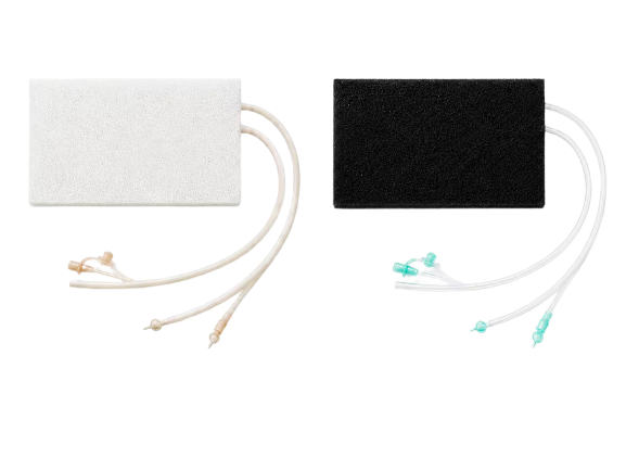
Select — Choose the appropriate VSD foam type: black polyurethane (PU) open-pore foam or white polyvinyl alcohol (PVA) foam, based on wound depth and exudate level.
Cut — Trim the foam to the exact wound shape using sterile scissors. Cut foam 1–2 cm larger than the wound margin to ensure full surface contact.
Pack — Fill every sinus, tunnel, and cavity completely. Zero dead space — no gaps or voids should remain anywhere in the wound bed.
Channel — Cut a side channel into the foam and embed the drainage tube 3–5 cm into the foam body to ensure even negative pressure distribution.
Protect — Apply a hydrocolloid or petroleum jelly strip around peri-wound skin to prevent direct foam contact with healthy tissue.
03Step 3 — Film Sealing
Protect Peri-Wound Skin — Apply a hydrocolloid dressing strip or petroleum jelly around the wound edge (3–5 cm margin) to prevent maceration from prolonged moisture exposure.
Drape Film — Cover the foam dressing and prepared skin margin with the transparent semi-permeable film. Work from centre outward, smoothing out all wrinkles to ensure full contact.
Seal Every Edge — Overlay film edges by at least 3–5 cm onto intact surrounding skin. Use additional film strips to reinforce all seams, overlaps, and tube exit sites.
Test for Leaks — Once negative pressure is applied, listen and feel around all margins for escaping air. Patch any whistling points immediately with an extra film patch.
Tube Exit Site — Create a small reinforced exit point 2–3 cm from wound edge, sealed with multiple film layers.
04Step 4 — Connect & Activate Negative Pressure
Connect — Attach the drainage tube securely to the VSD negative pressure device. Confirm all connections are tight and tube is free of kinks.
Set Pressure — Adjust to -125 to -450 mmHg, continuous or intermittent mode per clinician's order. For fresh post-operative wounds, start at -125 mmHg and increase gradually.
Observe Collapse — The foam should visibly collapse evenly and draw tightly against the wound bed. A visible reduction in foam volume confirms an effective sealed system.
Record Baseline — Document initial pressure setting, foam collapse status, drainage volume, and colour in the patient's clinical record.
Patient Briefing — Advise patient on precautions: avoid pulling the tube, report any unusual sounds, pain, or a sensation of wetness immediately.
05Step 5 — Monitor, Maintain & Dressing Change
Frequent Checks — Inspect every 2–4 hours: confirm pump is running, pressure is within set range, no alarms are active, and no air is escaping from the seal.
Monitor Drainage — Observe the collection canister: record volume, colour, and consistency (serous = normal; bloody = caution; purulent = infection). Alert clinician if drainage exceeds 500 mL/24h or suddenly increases.
Prevent Blockage — Never clamp the drainage tube. Keep tube free of kinks, twists, and dependent loops. If blockage is suspected, flush per protocol or notify the physician.
Dressing Change — Replace every 48–72 hours: turn off pump, carefully remove the film, inspect wound progress (granulation tissue, size reduction), gently re-debride if needed, and reapply fresh foam and film. For infected wounds, change every 24–48 hours.
Wean Off — Once wound has healthy granulation tissue coverage and dimensions are significantly reduced, transition to conventional dressing or reduce pressure per clinical decision.
💡 Click on any image to enlarge
Product Gallery
For: Ulcers · Burns · Donor sites · Pressure injuries (Stage I–II) · Post-dermatological procedure wounds
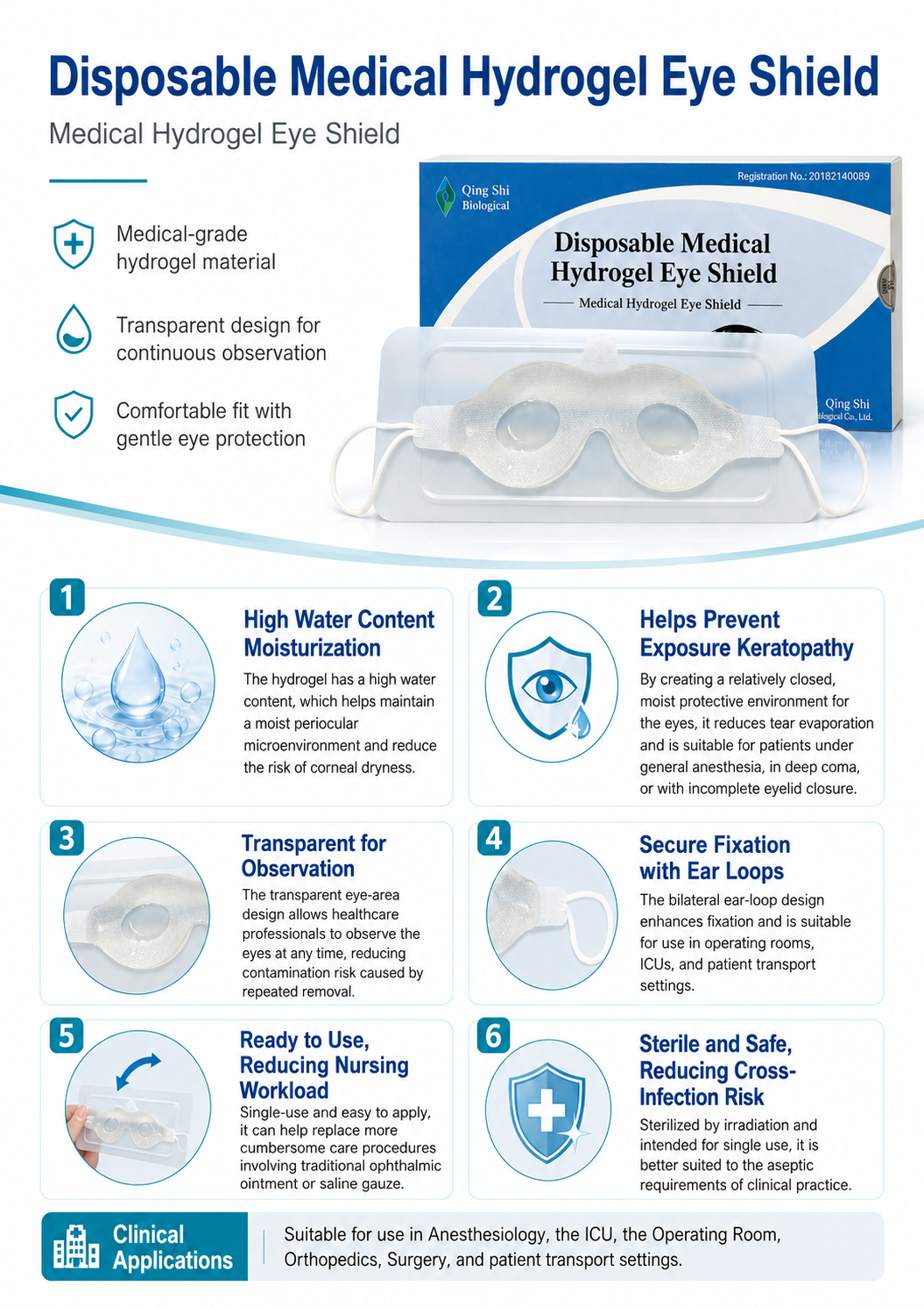
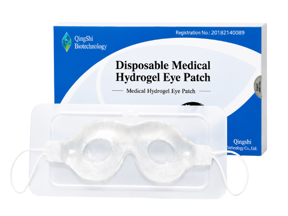
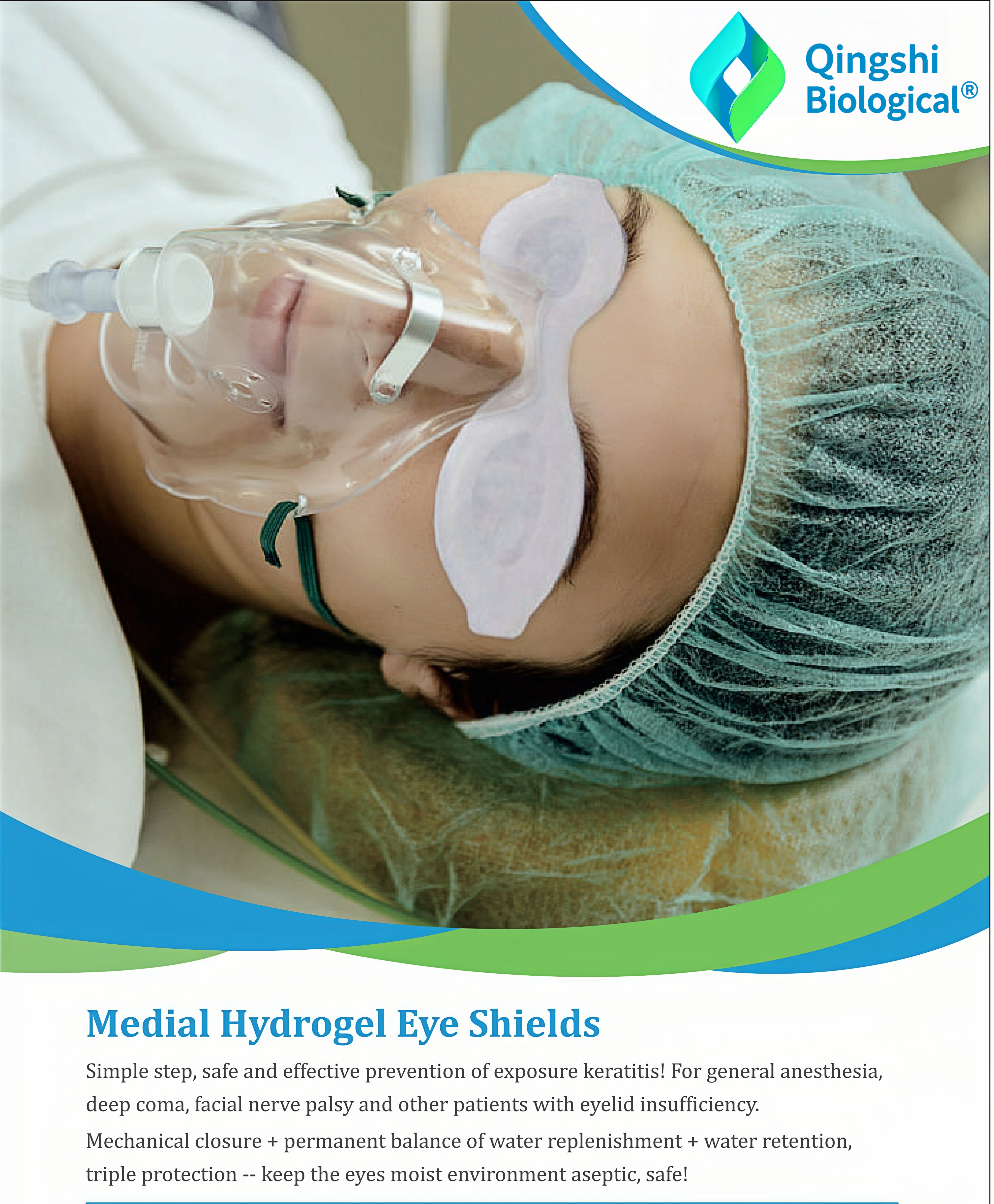
Request for QuoteFor: General anesthesia surgery · ICU long-term bedridden patients · Sedated emergency patients · Post-ophthalmic procedures
Request for QuoteQingshi Bio provides flexible cooperation models tailored to different market needs — enabling global partners to access high-quality wound care products with full technical and regulatory support.
Qingshi Bio allocates 100% of R&D capacity toward three interdependent domains: biomaterial science, wound microenvironment modulation, and controlled-delivery dressing systems — enabling clinical precision that generalist manufacturers cannot replicate.
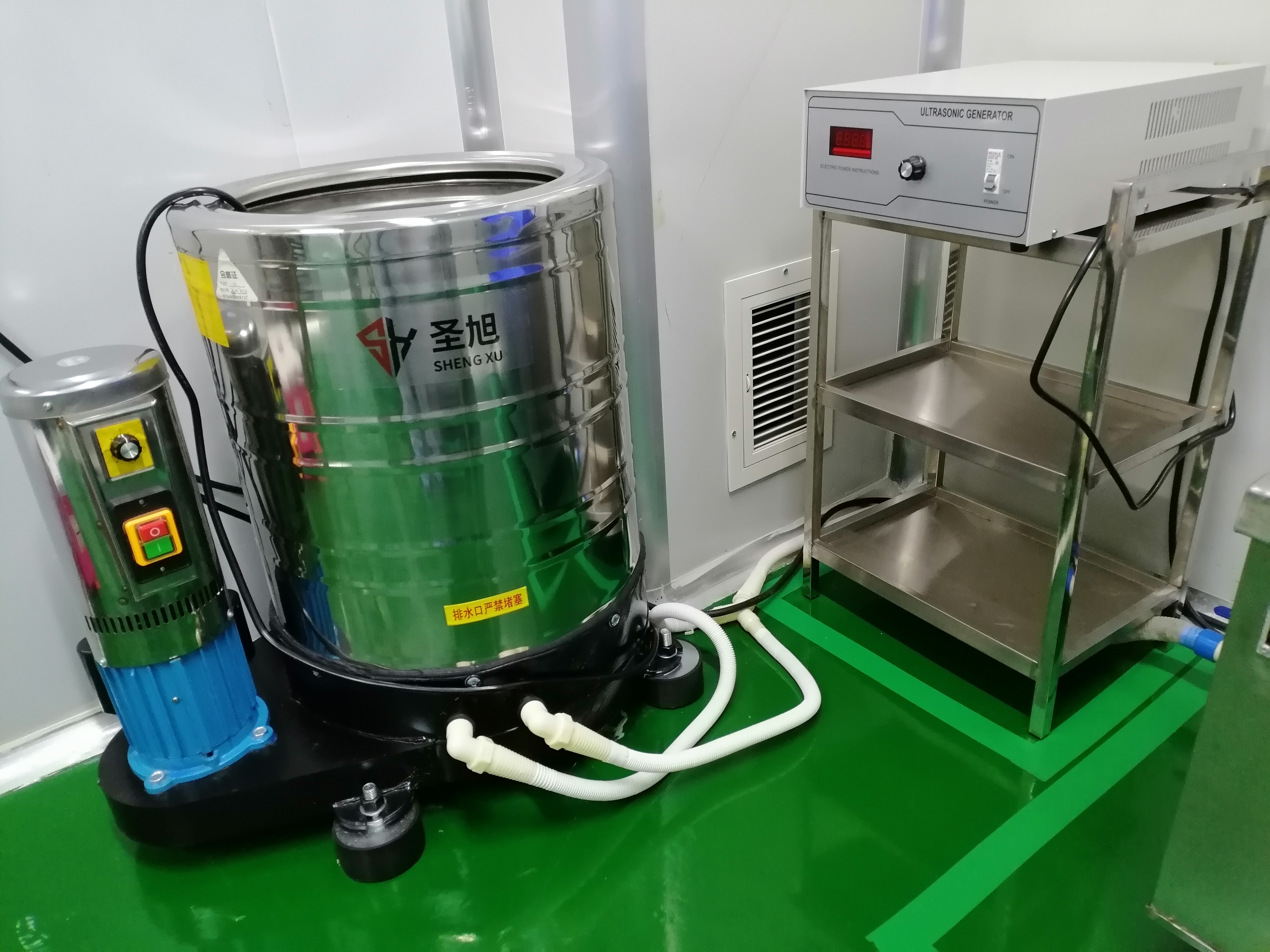
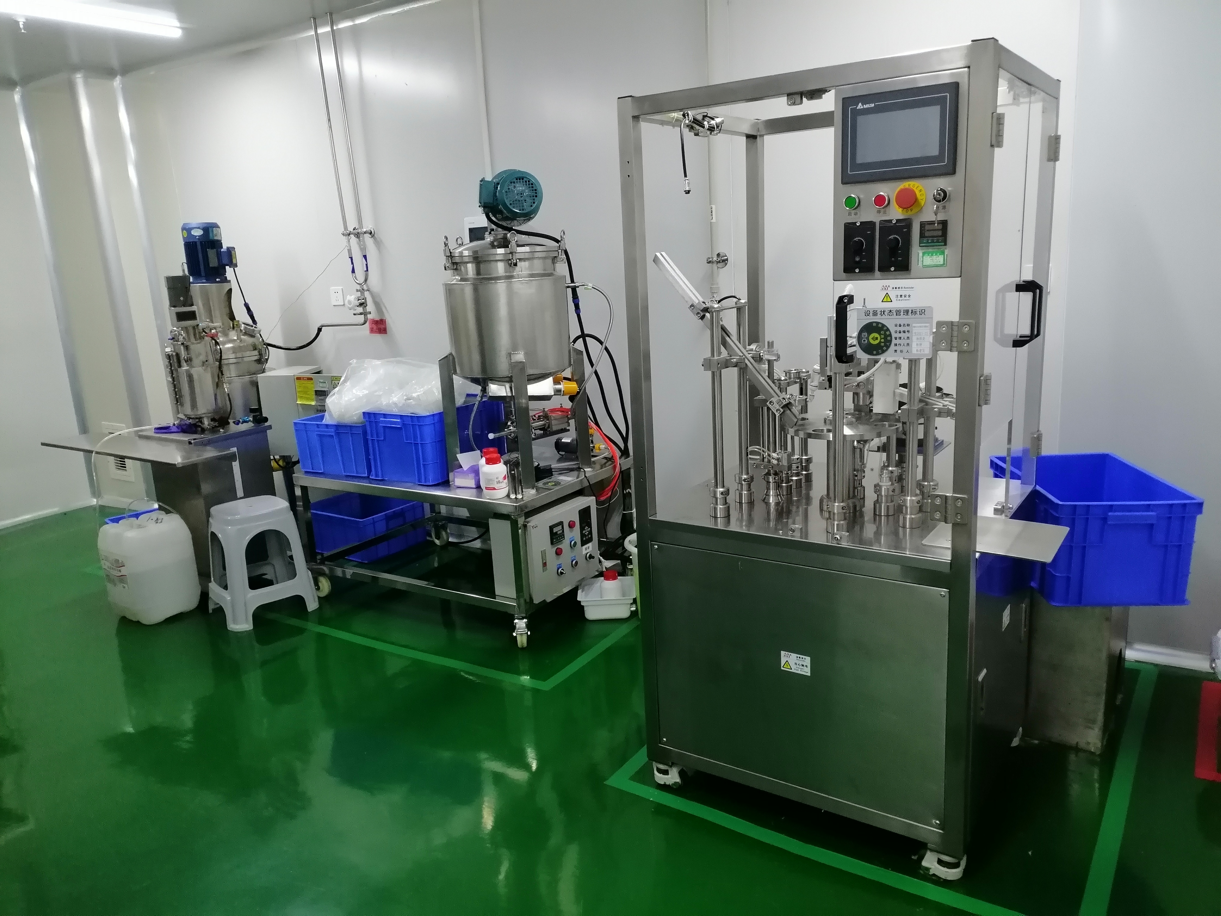
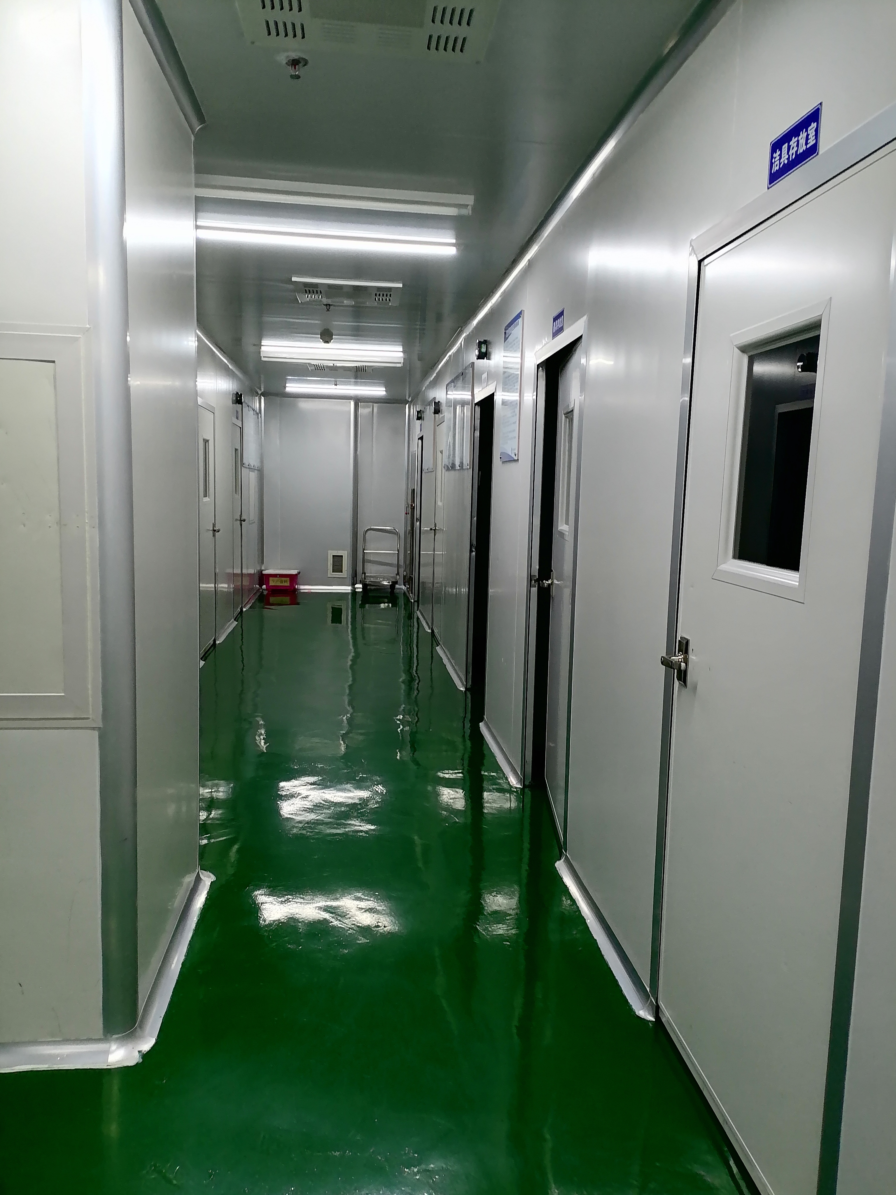
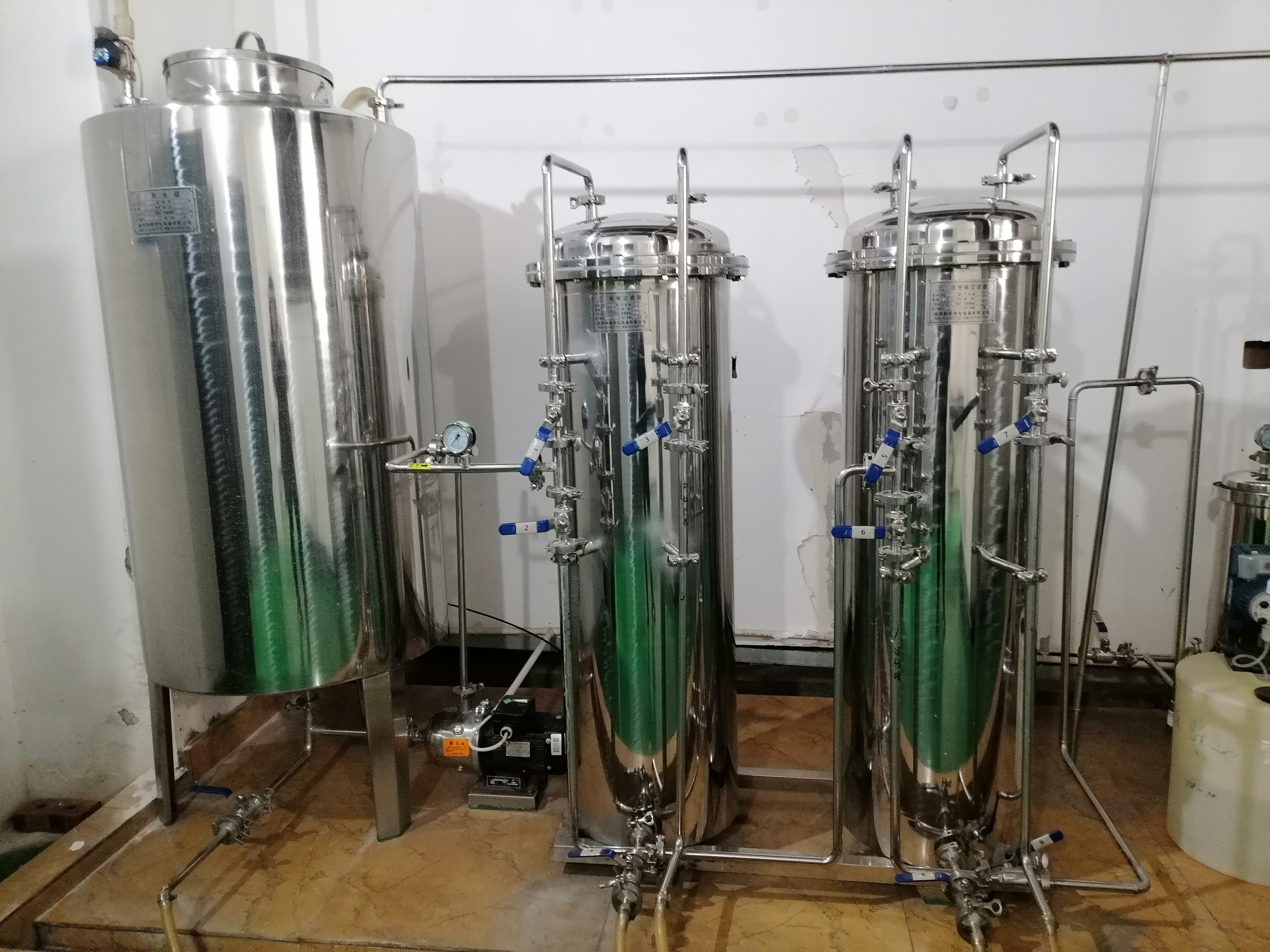
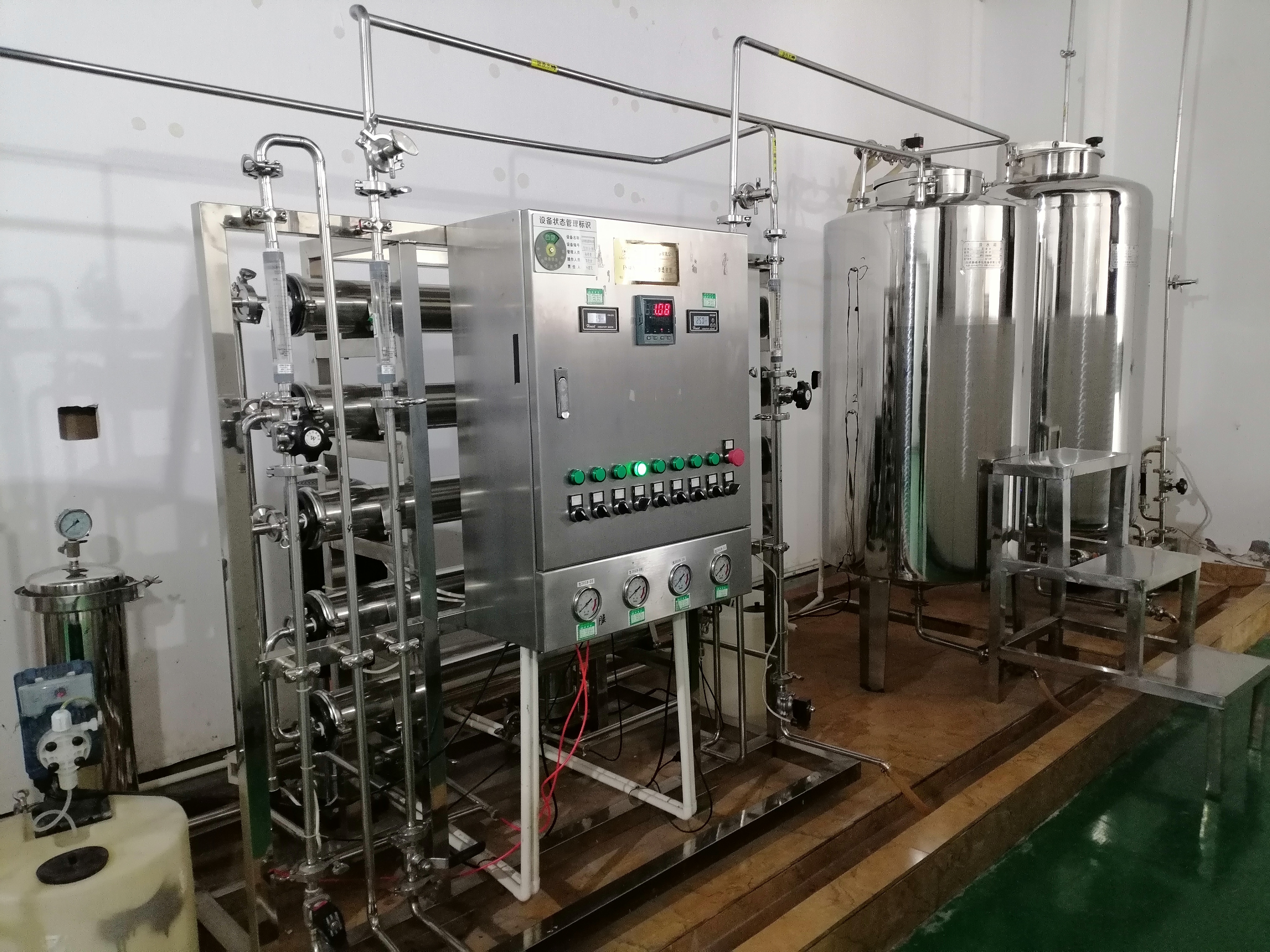
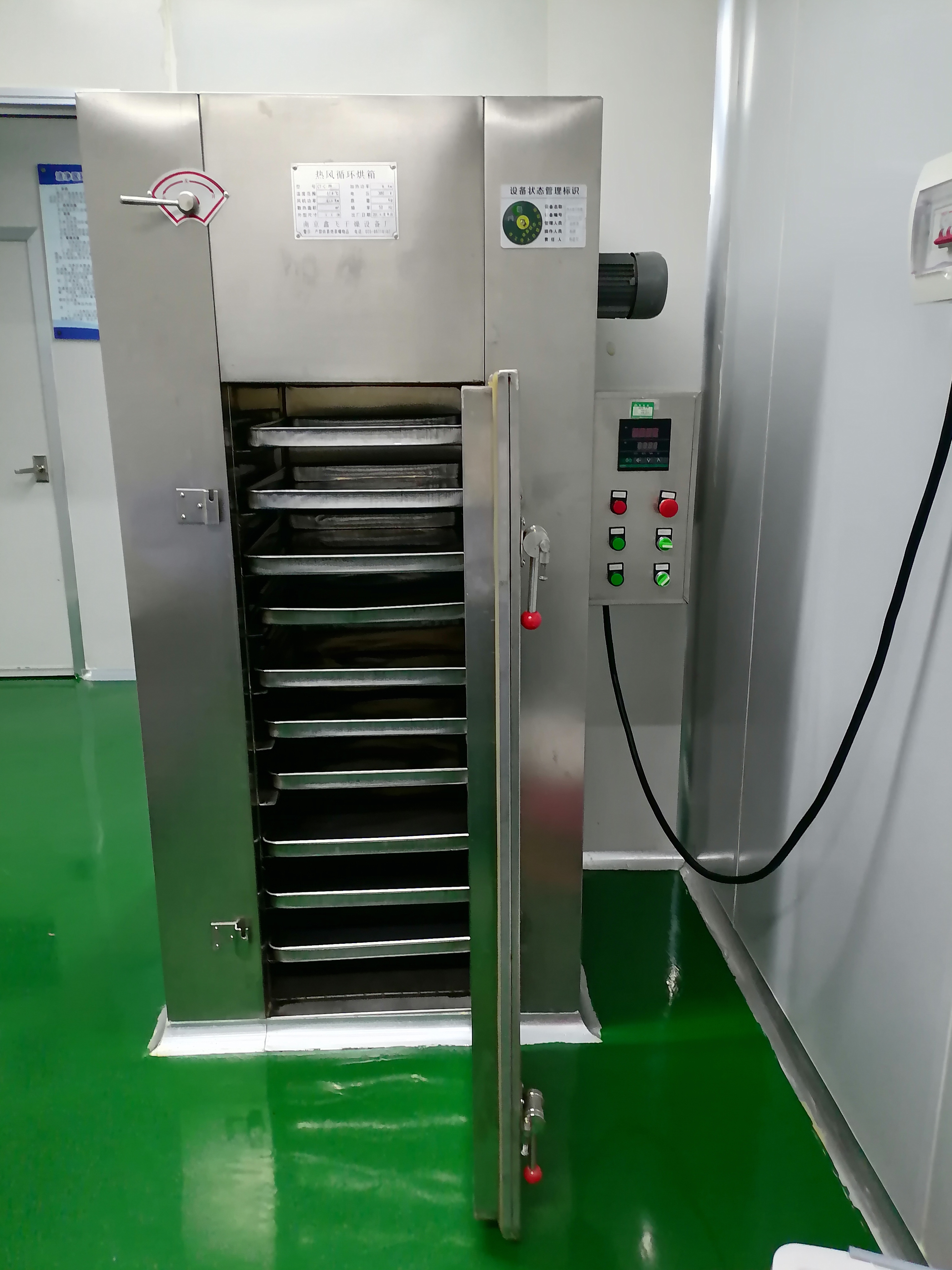
Fully integrated GMP-scale production facility — material compounding, precision molding, cleanroom assembly, and validated quality testing in a single site. No transfer risk. Lot-to-lot consistency guaranteed.
Qingshi Bio serves international markets through direct distributor partnerships, OEM/ODM collaboration, and private-label programs — with multi-language documentation and regulatory registration support.